Common names:
China |
Chinese Mandarin |
犬齿背眼虾虎鱼, 犬齒背眼鰕虎魚 (quan chi bei yan xia hu yu - canine-teethed dorsal-eyed goby), 中华钝牙虾虎鱼, 中華鈍牙鰕虎魚 (zhong hua dun ya xia hu yu - Chinese blunt-teethed goby), 中华尖牙虾虎鱼, 中華尖牙鰕虎魚 (zhong hua jian ya xia hu yu - Chinese sharp-teethed goby) |
Finland |
Finnish |
intiankitaryömijä |
Italy |
Italian |
saltafango coccodrillo* |
Viet Nam |
Vietnamese |
cá bống nheo |
United Kingdom |
English |
crocodile-face goby |
* proposed name
|
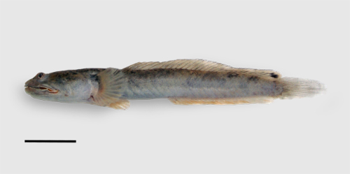
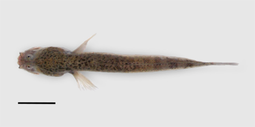
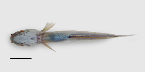
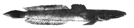
Oxuderces nexipinnis. Tanjung Piai, Johor, Peninsular Malaysia;
lateral view (above); dorsal view (centre); ventral view (below);
the bar is 10 mm long - freshly dead specimen (photo: G. Polgar, 2006)
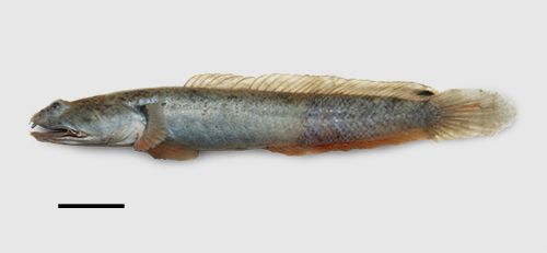
Oxuderces nexipinnis. Kukup Island, Johor, Peninsular Malaysia;
lateral view (right side, photo reversed);
the bar is 10 mm long
- freshly dead specimen (photo: G. Polgar, 2007)
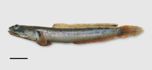
Oxuderces nexipinnis. Kukup Island, Johor, Peninsular Malaysia;
lateral view;
the bar is 10 mm long - anaesthetised specimen (photo: G. Polgar, 2007)
|
|
Synonyms:
Etymology:
'Oxuderces' comes from the Greek 'oxuderkês' (quick-sighted), which probably refers to the quick burying behaviour, elicited by visual
stimuli
'nexipinnis' means 'with connected fins' in Latin, which refers to the membrane connecting D1 and D2
|
Maximum recorded length:
81 mm SL (Jaafar & Parenti, 2016)
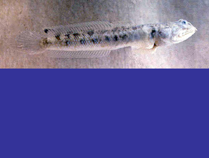
Live colouration (Murdy, 1989;
Takita et al., 1999; GP pers. obs.: Peninsular Malaysia):
ground colour dorsally greyish blue to pale brown, ventrally whitish to pale grey;
6 dark and irregular saddle-like bars and a series of dark blotches along lateral
midline may be visible; many irregular tiny brown speckles on nape and dorsum; shiny bluish scales can be present on flanks, on posterior half of trunk;
D1 translucent; D2 transparent except for a faint dusky medial stripe and a large
black ocellus with orange margin near distal tips of last 4 rays; caudal fin transparent to dusky or orange;
anal, pectoral and pelvic fins translucent, orange in some specimens;
large blackish blotch on upper base of pectoral fin; upper lip and tips of anterior nostrils lined with black;
upper lip posteriorly and ventrally orange. Eyes bright green to blue (see photo D)
Colouration on preservation
(Jaafar & Parenti, 2016; GP pers. obs.: Peninsular Malaysia):
ground colour pale to dark brown; dark spots, bars and blotches can be
retained, especially on head and nape; tips of anterior nostrils and upper lip lined with black or dark brown; dark brown blotch at the base of the pectoral fins;
all fins transparent except for the dusky ocellus at the posterior portion of D2
Diagnosis (Jaafar & Parenti, 2016):
first hemal spine extending ventrally to a point dorsal to mid-length of the first anal-fin pterygiophore (versus extending ventrally to
mid-length of the first anal-fin pterygiophore in O. dentatus); conspicuous
dermal invagination posterior to the point of attachment of pelvic-fin base; mouth superior; head length 27-30%SL.
The genus is diagnosed by (1) a fleshy external trough (deep groove or slit) running along the medial longitudinal axis in a dorsal interorbital
position (see photo G); the groove is supported internally by two ridges of the medially fused frontal bones (interorbital bridge) that curve towards
the midline, and is lined with thick epidermis and thin dermis; (2) highly thickened epidermis over the eye; (3) neural spine of the fourth vertebra broad, spatulate
and posteriorly directed; (4) portion of the anterior ceratohyal posterior to the insertion of the fourth branchiostegal ray elongate and notched (versus
not elongate, or elongate and not notched; see drawing D); and (5) the metapterygoid-symplectic-quadrate strut and the anguloarticular form an acute angle
Description (Jaafar & Parenti, 2016):
Head wider than deep: head length (HL) 27-30%SL, head width 33-44%HL, head depth 30-43%HL. Eye diameter 9-16%HL; interorbital distance 3-5%HL. Snout length 12-16%HL,
jaw length 41-60%HL. Body depth at anus 9-13%SL, body width at anus 4-7%SL. Predorsal length 28-32%SL. Length of base of D1 plus D2 55-59%SL; length of base of anal
fin 36-40%SL; pelvic-fin length 14-18%SL; pectoral-fin length 14-19%SL; caudal-fin length 21-25%SL. D2 with 25-26 elements; anal fin with 24-26 elements; pectoral rays 21-24.
Lateral longitudinal scale count 50-54; ventral region of head, cheek, operculum, isthmus, dorsal region anterior to D1 and pectoral-fin bases scaleless, except in largest specimens;
all scales small and embedded.
All congeners also have: depressed anterior portion of the head and pointed snout profile. Distinct notch in middle of upper lip between two
medial premaxillary teeth; thick lips, with posterior lip distally protruding. Gape wide, extending 3-4 eye lengths posterior to the orbit; preopercle thin and
crescent-shaped, five thin branchiostegal rays. Roof of mouth with fleshy ellyptical palp with pointed tips, studded with papillae. Oral jaw teeth in single
row both on premaxilla and dentary, all caninoid; premaxillary teeth decreasing in length posteriorly, dentary teeth more uniform in size; 1-2 canine teeth
on both sides of premaxillary symphysis, longer than other teeth, extending anteroventrally and projecting beyond lower jaw when mouth closed; no canine teeth
on both sides of mandibular symphysis; no vomerine and palatine teeth. Gill opening restricted, beginning from a point anterior to midpoint of pectoral-fin
base, then coursing anteroventrally to a point dorsal to the pelvic fin origin. Eyes not meeting medially, without dermal cup. Supraorbital pore C in the
anterior portion of the fleshy external interorbital trough; anterior oculoscapular pores; large posterior nostril, anteroventral to eye; anterior nostril
at tip of pendulous short tube overlapping lower jaw. D1 and D2 connected by membrane for entire height; D2 or anal fin and caudal fin not connected by membrane;
D1 with six spines; all elements of D2 and anal-fin segmented rays, last two rays sharing same pterygiophore; pelvic fins fused in a pelvic disc, not reaching
genital papilla; caudal fin lanceolate with 17 segmented rays; dorsal procurrent rays 5; ventral procurrent rays 4; 2 epural bones. Male genital papilla triangular
and conical, with posterior tip pointed; female genital papilla rectangular and bulbous
Diet:
no published study is available
Reproduction:
no published study is available
|
Ecological notes (Polgar & Bartolino, 2010: as
O. dentatus):
locally abundant on lower mudflats, moving in areas of mud covered by a thin film of water;
larger individuals (>7-8 cm) can be caught by trawl nets on lower mud flats at high tide
(Murdy, 1989; GP pers. obs.: Peninsular Malaysia). O. nexipinnis stores air in burrows
(Ishimatsu et al., 2000). In Malaysia, it is a common prey of homalopsid snakes
(Bitia hydroides; Jayne et al. 1995; and Cerberus rynchops;
Jayne et al. 1988). O. nexipinnis can be ecologically partitioned between larger individuals, more abundant at lower levels of the mudflat, and smaller ones, more abundant at higher levels;
a similar pattern was observed in different habitats of the same ecosystem in
Boleophthalmus boddarti and Periophthalmus gracilis,
while P. variabilis apparently exhibited an inverse pattern
(Polgar & Bartolino, 2010)
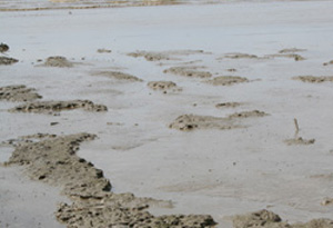
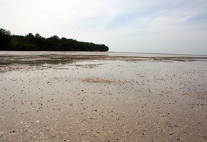
left: Tanjung Piai, Peninsular Malaysia: lower mudflat at low tide; Ox.
nexipinnis was here abundant
right: Kukup Island, Peninsular Malaysia: a shot of the mangrove forest and of the adjacent tidal flat at low tide; the mud layer was less than 30 cm deep, with shell
lag deposits (Placuna sp.) beneath
(photo: G. Polgar, 2006; 2007)
|
|
|
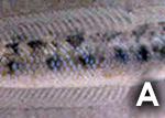
A: freshly dead specimen (photo: E.O. Murdy, Muar, Peninsular Malaysia,
1985);
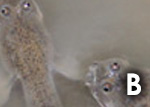
B: two specimens in aquarium (photo: G. Polgar; collected in Tanjung Piai, Peninsular Malaysia, 2006);
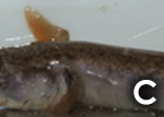
C: in aquarium, out of water (photo: G. Polgar, collected in Kukup Island, Peninsular Malaysia, 2007);
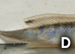
D: an underwater shot of the specimen in C (photo: G. Polgar, 2007);
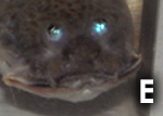
E: another close-up of the same individual (photo: G. Polgar, 2007);
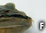
F: detail of the diagnostic dermal invagination (red arrow) posterior to the point of attachment of
pelvic-fin base (ZRC 54696, West Bengal,
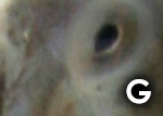
India; photo: G. Polgar, 2017); G: dorsal view of the interorbital trough (T) diagnostic for Oxuderces spp., and of the
position of the supraorbital pore C; black scale bar = 1 mm (photo: G. Polgar, 2007; freshly dead specimen collected in Kukup Island, Peninsular Malaysia) - * with permission from the author

|
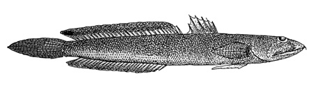
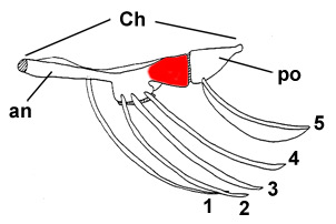
Drawings of Oxuderces nexipinnis:
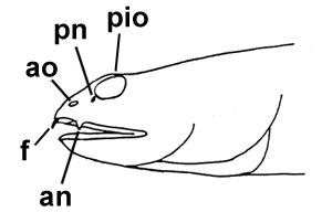
A: cephalic sensory and nasal pores of Oxuderces spp.: an = anterior nostril;
ao = anterior oculoscapular canal pore; f = caninoid teeth; pio = posterior interorbital pore; pn = posterior nostril
(modified from Murdy, 1989)*; B: Apocryptichthys cantoris (Day, 1871) (Day, 1889, Fig. 94; illustration of a specimen from Madras or Andamans); C: Apocryptichthys cantoris (Day, 1871) (Koumans, 1953); D: lateral view
of the hyoid arch of Ox. dentatus, rotated about 45° clockwise; the portion of the anterior ceratohyal that is not present
in other oxudercine genera is coloured in red: Ch = ceratohyal; an = anterior ceratohyal; po =
posterior ceratohyal; 1-5 = branchiostegals (modified from Murdy, 1989)* - * with permission
|